Clinical
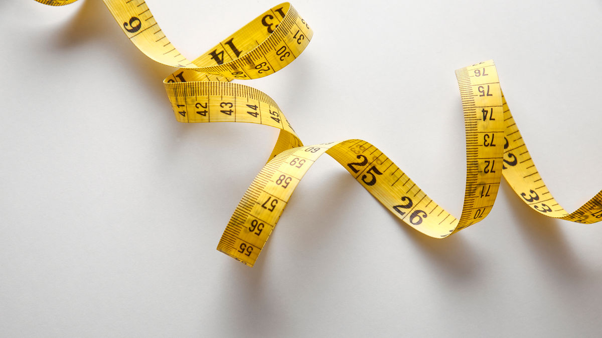
Axial length measurement in myopia management - how often and how much change is normal?
Sponsored by
In this article:
How frequently should we measure axial length in myopia management practice, and how should it best direct our treatment strategy? Colleagues raised questions about this in the Myopia Profile Facebook group (link) - here we discuss how axial length change is related to refraction and ethnicity, and how to determine whether an axial length change is normal due to emmetropization or indicating myopia progression.
How frequently should we measure axial length?
Most commenters suggest monitoring myopic patients every 6 months or to see them more often if the patient is a faster progressor.
There were also discussions about how to determine the normal, expected amount of axial length change in children due to emmetropization, and separating this normal growth from myopia progression growth.
Jones et al also shows that the corneal curvature of myopes tends to stay stable whilst emmetropes’ flatten over time. An emmetropes’ cornea flattens while axial length increases, maintaining a balance in emmetropia. Meanwhile, a myope’s cornea does not change in curvature with axial length change, leading to an increase in myopic refractive error.1 It’s not all about the cornea, though – the crystalline lens likely plays a bigger role in the loss of balance in refractive components that occurs in myopia development. Mutti and colleagues2 found that in children who became myopic, before onset the crystalline lens underwent similar thinning, flattening and loss of power to children who stayed emmetropic. Within a year of myopia onset, though, the crystalline lens stopped these compensatory changes which continue in emmetropes throughout normal childhood axial elongation. Also, it is important to remember that some component of childhood axial length increases are not indicative of myopia progression, and are in fact normal.
If you wish to compare the patient’s axial length change to the expected axial length change of an emmetrope, this is possible using a formula derived from the growth curve of emmetrope as TA suggested.1 This will allow a practitioner to differentiate abnormal axial length change related to myopia from normal physiological axial length growth. This model only applies to children less than 10.5 years old. A different model applies to those more than 10.5 years old: =21.353 + 0.759 x ln(age). See reference 1 below for more info.
As described in Six questions on axial length in myopia management the refraction-to-axial-length ratio is variable across age groups, studies and the range of both measurements so isn’t so easy to simply define. A big part of the complexity is due to the variability in measurement of both refraction and axial length. Here are two exampes from multifocal contact lens myopia control studies. In the MiSight three year study,3 the 1mm = 2.40D in both their treatment and control groups. In the newly published BLINK study,4 1mm = 1.4 to 1.6D across the treatment and control groups. This is despite similar age ranges, ethnicities and methologies in both studies. The simple answer? We’re still learning.
The table below shows the percentiles of axial elongation European and Chinese children reported by Diez PS et al and Tideman JW et al.5,6 This shows that children of Chinese descent have longer axial lengths than those of European descent.
| Percentile | Female | Male | |||
| European | Chinese | European | Chinese | ||
| 6 years | 25 | 21.66 | 22.03 | 22.14 | 22.55 |
| 50 | 22.06 | 22.54 | 22.59 | 22.99 | |
| 75 | 22.49 | 23.04 | 23.01 | 23.50 | |
| 9 years | 25 | 22.33 | 23.16 | 22.83 | 23.70 |
| 50 | 22.79 | 23.72 | 23.31 | 24.32 | |
| 75 | 23.25 | 24.31 | 23.79 | 24.89 | |
| 15 years | 25 | 22.68 | 23.83 | 23.17 | 24.39 |
| 50 | 23.15 | 24.37 | 23.65 | 25.01 | |
| 75 | 23.65 | 25.20 | 24.21 | 25.80 | |
How much change in axial length is required for a change in myopia control strategy?
There is no definite value suggested for one to change the strategy. Given the variability in axial length change based on age and ethnicity, the best approach is to change strategy if the current one does not sufficiently reduce myopia progression. This can be gauged when progression is higher than that expected as reported in the literature.
Axial length progression can be gauged using the percentile charts above - a child who jumps to a higher percentile over time is likely demonstrating accelerated growth.
To use another example from the literature, the COMET progressive addition spectacle lens trial analysed a variety of factors for their influence on myopia progression and found the strongest relationship was with baseline age. The progressive addition lens (PAL) intervention showed only a small treatment effect over three years. Axial length elongation by age was evaluated in an ethnically diverse group (46% White, 26% African American, 15% Hispanic, 8% Asian, 5% Mixed).7
Firstly, annual increases in overall axial length were 0.28 mm (± 0.01) between baseline and 1-year visit (at which time the children were 10.3 years on average); 0.21 mm (± 0.008) between 1 and 2 years (by age 11.3 years) and 0.17 mm (± 0.01) between 2 and 3 years (by age 12.3 years), reflecting the slowing in eye growth over time.
Secondly, when results were pooled for all children (treatment PAL and control SV spectacle groups), unadjusted axial elongation over three years was:
- 1.08mm in children who were 6-7 years old at baseline
- 0.82mm in 8 year olds
- 0.68mm in 9 year olds
- 0.57mm in 10 year olds
- 0.45mm in 11 year olds.
To determine if a treatment strategy is working as expected, one would compare the axial elongation likely in a single vision corrected myopic child - as provided above - to the expected percentage reduction with the strategy. For more help on understanding efficacy you can read this blog, and also get more help on gauging success in this blog.
Take home messages:
- To monitor axial length change, the general consensus suggests 6-monthly measurements or more frequently in rapid progressors.
- We can monitor axial length change by comparing to the normative value expected for the patient’s age. If your patient’s result falls outside of normal limits, then it is worthwhile considering a more comprehensive myopia control strategy.
- Chinese children tend to have longer axial lengths and experience faster axial elongation than European children, even in normal emmetropic eye growth.
- The rate of axial elongation varies in different age groups, with younger children increasing the fastest.
- Using axial length as a gauge of myopia management success is still a little difficult, but models exist as shown above. More research findings and development of growth charts for larger populations including a variety of ethnicities will help to answer this question.
Further reading:
Meet the Authors:

About Connie Gan
Connie is a clinical optometrist from Kedah, Malaysia, who provides comprehensive vision care for children and runs the myopia management service in her clinical practice.
Read Connie's work in many of the case studies published on MyopiaProfile.com. Connie also manages our Myopia Profile and My Kids Vision Instagram and My Kids Vision Facebook platforms.

About Kimberley Ngu
Kimberley is a clinical optometrist from Perth, Australia, with experience in patient education programs, having practiced in both Australia and Singapore.
Read Kimberley's work in many of the case studies published on MyopiaProfile.com. Kimberley also manages our Myopia Profile and My Kids Vision Instagram and My Kids Vision Facebook platforms.
This content is brought to you thanks to an educational grant from
References
- Jones LA, Mitchell L, Mutti DO, Hayes JR, Moeschberger ML, Zadnik K. Comparison of ocular component growth curves among refractive error groups in children. Clinical and Epidemiologic Research. 2005;46:2317-2327. (link)
- Mutti DO, Mitchell GL, Sinnott LT, et al. Corneal and crystalline lens dimensions before and after myopia onset. Optom Vis Sci. 2012;89(3):251-262. (link)
- Chamberlain P, Peixoto-de-Matos SC, Logan NS, Ngo C, Jones D, Young G. A 3-year randomized clinical trial of MiSight lenses for myopia control. Optom Vis Sci. 2019;96:556-567. (link)
- Walline JJ, Walker MK, Mutti DO, et al. Effect of High Add Power, Medium Add Power, or Single-Vision Contact Lenses on Myopia Progression in Children: The BLINK Randomized Clinical Trial. JAMA. 2020;324:571–580. (link)
- Diez PS, Yang LH, Lu MX, Wahl S, Ohlendorf A. Growth curves of myopia-related parameters to clinically monitor the refractive development in Chinese schoolchildren. Graefe's Arch Clin Exp Ophthalmol. 2019;257:1045-1053. (link)
- Tideman JW, Polling JR, Vingerling JR, Jaddoe VW, Williams C, Guggenheim JA, Klaver CC. Axial length growth and the risk of developing myopia in European children. Acta ophthalmologica. 2018;96:301-309. (link)
- Hyman L, Gwiazda J, Hussein M, Norton TT, Wang Y, Marsh-Tootle W, Everett D. Relationship of age, sex, and ethnicity with myopia progression and axial elongation in the correction of myopia evaluation trial. Arch Ophthalmol. 2005;123:977-987. (link)
Enormous thanks to our visionary sponsors
Myopia Profile’s growth into a world leading platform has been made possible through the support of our visionary sponsors, who share our mission to improve children’s vision care worldwide. Click on their logos to learn about how these companies are innovating and developing resources with us to support you in managing your patients with myopia.