Clinical
Which clinical tests are required for myopia management?
Sponsored by
If you are just getting started in your myopia treatment journey, or are keen to check in on best-practice processes, the following clinical article will take you through advice from the International Myopia Institute (IMI) - a panel of global experts. The IMI has published two landmark volumes of white paper reports on all aspects of myopia, the first in 2019 and the second in 2021. The foundation to the advice provided below will be the IMI Clinical Management Guidelines report,1 with references to several more. You can review Volume 1 and Volume 2 White Paper Reports and read the Clinical Summaries (available in several languages) via this link.
In this article:
History
The case history for myopia management serves to understand your patient's risks for myopia onset or progression and suitability for myopia control treatments. Once a child is myopic, the biggest risk factor for progression to high myopia is age of onset,2 and when combined with a recent history of fast progression (more than 1D in a year) helps to predict faster subsequent progression in children under age 10.3 Determining age of onset and prior progression history is helpful to formulating how proactive your myopia management strategy should be.
Additional risk factors for myopia onset or progression are family history, with more myopic parents and higher parental myopia increasing risk.4 Questions on ocular and general health, and previous myopia corrections and treatments, will also help to guide your recommendations.
Finally, since the IMI Risk Factors for Myopia Report explains that outdoor time and near work habits are linked to myopia development and progression,5 asking these questions in the case history will help to lay the foundation of the eventual discussion about managing the visual environment. It will also help you to determine visual tasks, sports and hobbies to guide treatment selection, particularly the best optical treatment.
Tests to do: ask about age of myopia onset, prior myopia progression (if available), family history, ocular and general health, previous myopia corrections and/or treatments, outdoor time and near work habits, sports and hobbies.
Refraction and acuity
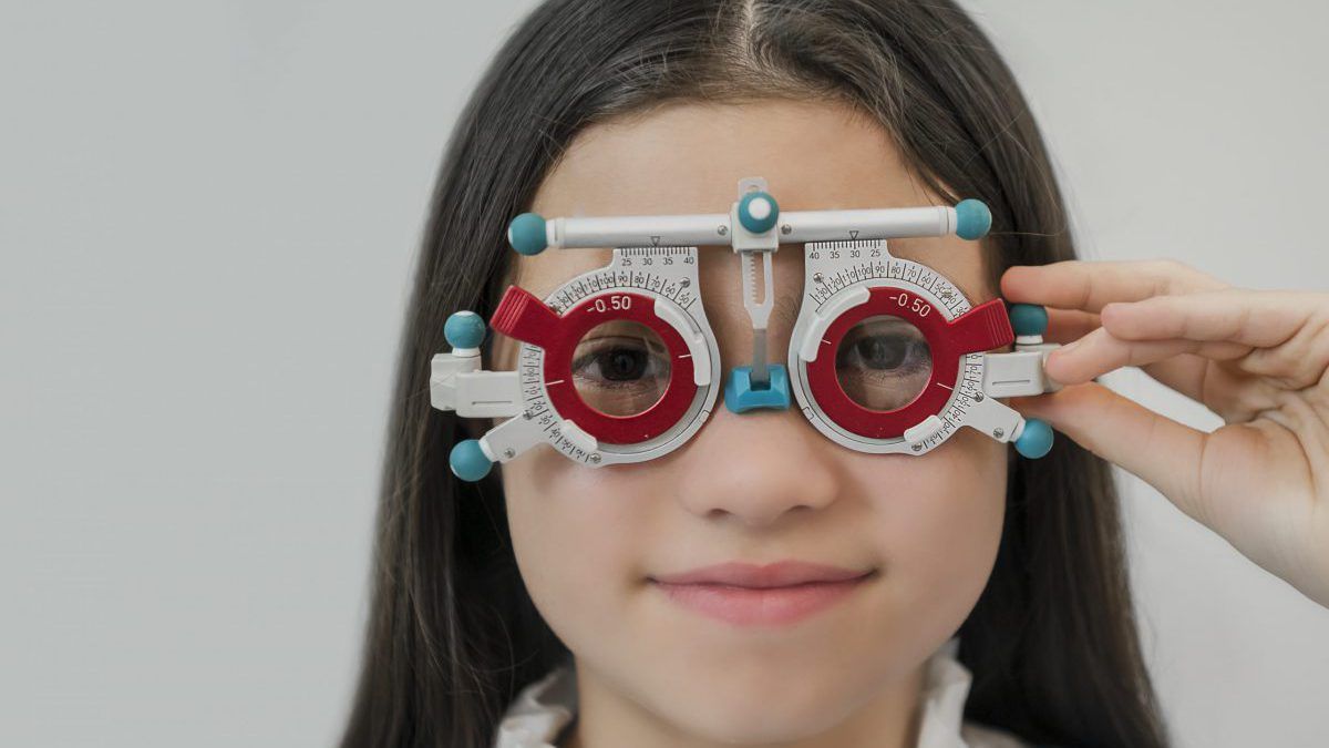
The most important element of the examination is accurately defining the level of myopia. Measuring acuity with the current correction and after updated refraction is important clinically, as well as for parents to understand the functional impact of myopia progression.
The IMI doesn’t specify a preferred method of refraction, but retinoscopy is an excellent screening tool for detecting refractive error in children.6
The IMI Clinical Management Guidelines1 suggest using cycloplegia when indicated, which may vary depending on practitioner, country, availability and the presentation of the patient. The IMI defines myopia by refraction as: "when ocular accommodation is relaxed. These definitions avoid the requirement for objective refraction so as to be independent of technique, but by making reference to relaxation of accommodation are compatible with both cycloplegic and standard clinical subjective techniques." If used, the recommended dosage for cycloplegic refraction is two drops of 1% tropicamide or cyclopentolate given 5 minutes apart. Cycloplegic refraction should be performed 30 to 45 minutes after the first drop is instilled.
There is more guidance on measuring acuity and refraction in children of all ages, including how to refract if you don't have access to cycloplegia, in our article How to Achieve Accurate Refractions for Children.
Tests to do: the most accurate measure of refraction possible, with acuity measured before and after refraction.
Binocular Vision and Accommodative Tests
Binocular vision disorders in children have been linked to earlier onset, faster progression and different outcomes in the treatment of myopia. The IMI Clinical Management Guidelines1 state that there is no gold standard binocular vision assessment process. It recommends "tests that assess the various elements of accommodation and vergence...[and] the same tests need to be used in follow-up consultations to monitor for changes." Most of these tests are measures of near point function. The recently published IMI Accommodation and Binocular Vision in Myopia Development and Progression Report indicates that the most common measures in clinical studies are accommodative lag, near phoria and accommodation-convergence (AC/A) ratio.7
Here are links to Myopia Profile technique articles and videos to help you learn more on how to assess:
- Accommodative lag,
- Accommodative facility,
- Near phoria and
- Fusional vergence reserves at near.
- To understand how and why binocular vision disorders are relevant to myopia onset, progression, treatment response and even the newest research, read Four reasons why binocular vision matters in myopia management.
You can also read Getting Started In Myopia Management: What Equipment Do I Need? for more detail on the specific tests and equipment utilized in clinical studies of myopia and binocular vision.
Tests to do: measure accommodation and vergence function, with a focus on near point function - especially accommodative lag, near phoria and AC/A ratio.
Anterior eye health evaluation
As part of a thorough paediatric eye examination, it is important to assess the health of the anterior eye, especially if considering contact lens fitting. The IMI Clinical Management Guidelines also recommend measuring intraocular pressure (IOP). This can be challenging in young children and may not be possible for some eye care practitioners based on scope of practice - you will know best what comprises suitable clinical care pathways in your country.
Corneal Topography
Corneal topography is indicated when contact lenses are going to form part of the management plan. Orthokeratology is only possible with good topography scans. Topography is also indicated if there are corneal signs, astigmatism or unusual retinoscopy reflexes. Some topographers also have dry eye assessment and management suites.
Tests to do: anterior eye health evaluation. IOP measurement and corneal topography measurement where indicated and/or possible.
Posterior eye health evaluation
The IMI recommends retinal health examination in all myopes, and annually in high myopes through dilated pupils as indicated. Additional imaging with fundus photos and/or OCT is recommended if retinal findings are noted, or to objectively document retinal features.1
The newly published IMI Pathologic Myopia report8 explained that while high myopia is typically defined as more than 5D or axial length more than 26.5mm, pathologic myopia is defined by the presence of posterior staphyloma or myopic maculopathy and can occur in eyes without high myopia. Advanced imaging and/or co-management with ophthalmology is prudent in these cases.
Axial Length
The popularity of axial length as a tool to monitor myopia control outcomes is growing, yet it is still not widespread in many eye care practices. This is partly due to access and cost issues; and partly due to the science still establishing how to best utilize the metrics. New technological innovations are addressing the former, whilst understanding on typical emmetropic eye growth and percentile growth charts are addressing the latter.
Axial length is useful as a single measure to indicate myopia-associated eye disease risk and as a repeated measure to gauge myopia management outcomes. Read more in the related article Getting Started In Myopia Management: What Equipment Do I Need? Ideally this should be measured every six months, but if you don't have access to the equipment, consider if you could work with a colleague optometrist or ophthalmologist for annual measurements. If you are an optometrist without access to mydriatic diagnostic drugs, depending on your scope of practice and appropriate clinical pathways in your country, you may also need to co-manage with ophthalmology for annual retinal health examination through dilated pupils for high myopes, and other myopes on indication.
Tests to do: retinal health examination and axial length measurement done at least annually, or on indication. Consider co-management to provide the best possible care.
You have everything you need to get started
The baseline and follow up examinations for myopia management encompass standard primary eye care processes. Whilst scope of practice varies across the world for the eye care professions, this shouldn't be a barrier to myopia management - if you don't have access to cycloplegic refraction, mydriatic retinal health examination or measurement of axial length, there are options to alter clinical techniques or consider co-management. The time to start is now!
Further reading on getting started in myopia management
Meet the Authors:
About Cassandra Haines
Cassandra Haines is a clinical optometrist, researcher and writer with a background in policy and advocacy from Adelaide, Australia. She has a keen interest in children's vision and myopia control.
This content is brought to you thanks to an educational grant from
References
- Gifford KL, Richdale K, Kang P, Aller TA, Lam CS, Liu YM, Michaud L, Mulder J, Orr JB, Rose KA, Saunders KJ, Seidel D, Tideman JWL, Sankaridurg P. IMI - Clinical Management Guidelines Report. Invest Ophthalmol Vis Sci. 2019 Feb 28;60(3):M184-M203. (link)
- Chua SY, Sabanayagam C, Cheung YB, Chia A, Valenzuela RK, Tan D, Wong TY, Cheng CY, Saw SM. Age of onset of myopia predicts risk of high myopia in later childhood in myopic Singapore children. Ophthalmic Physiol Opt. 2016 Jul;36(4):388-94. (link)
- Matsumura S, Lanca C, Htoon HM, Brennan N, Tan CS, Kathrani B, Chia A, Tan D, Sabanayagam C, Saw SM. Annual Myopia Progression and Subsequent 2-Year Myopia Progression in Singaporean Children. Transl Vis Sci Technol. 2020 Dec 7;9(13):12. doi: 10.1167/tvst.9.13.12. PMID: 33344056; PMCID: PMC7726587. (link)
- Jiang X, Tarczy-Hornoch K, Cotter SA, et al. Association of Parental Myopia With Higher Risk of Myopia Among Multiethnic Children Before School Age. JAMA Ophthalmol. 2020;138(5):501-509. (link)
- Morgan IG, Wu PC, Ostrin LA, Tideman JWL, Yam JC, Lan W, Baraas RC, He X, Sankaridurg P, Saw SM, French AN, Rose KA, Guggenheim JA. IMI Risk Factors for Myopia. Invest Ophthalmol Vis Sci. 2021 Apr 28;62(5):3. (link)
- Schmidt P, Maguire M, Dobson V, Quinn G, Ciner E, Cyert L, Kulp MT, Moore B, Orel-Bixler D, Redford M, Ying GS; Vision in Preschoolers Study Group. Comparison of preschool vision screening tests as administered by licensed eye care professionals in the Vision In Preschoolers Study. Ophthalmology. 2004 Apr;111(4):637-50. (link)
- Logan NS, Radhakrishnan H, Cruickshank FE, Allen PM, Bandela PK, Davies LN, Hasebe S, Khanal S, Schmid KL, Vera-Diaz FA, Wolffsohn JS. IMI Accommodation and Binocular Vision in Myopia Development and Progression. Invest Ophthalmol Vis Sci. 2021 Apr 28;62(5):4. (link)
- Ohno-Matsui K, Wu PC, Yamashiro K, Vutipongsatorn K, Fang Y, Cheung CMG, Lai TYY, Ikuno Y, Cohen SY, Gaudric A, Jonas JB. IMI Pathologic Myopia. Invest Ophthalmol Vis Sci. 2021 Apr 28;62(5):5. (link)
Enormous thanks to our visionary sponsors
Myopia Profile’s growth into a world leading platform has been made possible through the support of our visionary sponsors, who share our mission to improve children’s vision care worldwide. Click on their logos to learn about how these companies are innovating and developing resources with us to support you in managing your patients with myopia.

