Science
The Menicon IMC 2024 showcase: ortho-k efficacy, safety and ocular changes

Sponsored by
In this article:
The article provides an overview of Menicon’s contributions to the International Myopia Conference 2024, highlighting their research in ortho-k and its efficacy, safety, and effects on ocular structures.
- Eleven years of orthokeratology contact lens wear for slowing myopia progression in children
- A novel approach for estimating the efficacy of orthokeratology lenses in slowing myopia progression
- Comparison of axial elongation rates before, during, and after orthokeratology
- Cleaning efficacy and anti-bacterial properties of contact lens care solutions for orthokeratology lenses
- Effect of coexistence with bacteria treated with multipurpose solution on excystment of Acanthamoeba castellanii
- Effect of ortho-k lenses on cornea endothelial cells, cornea thickness, and ocular surface in myopic children
- Comparison of peripheral eye length measurements between Orthokeratology and single vision spectacle lens wearers, using MRI analysis
- Factors associated with axial length and subfoveal choroidal thickness in myopic children
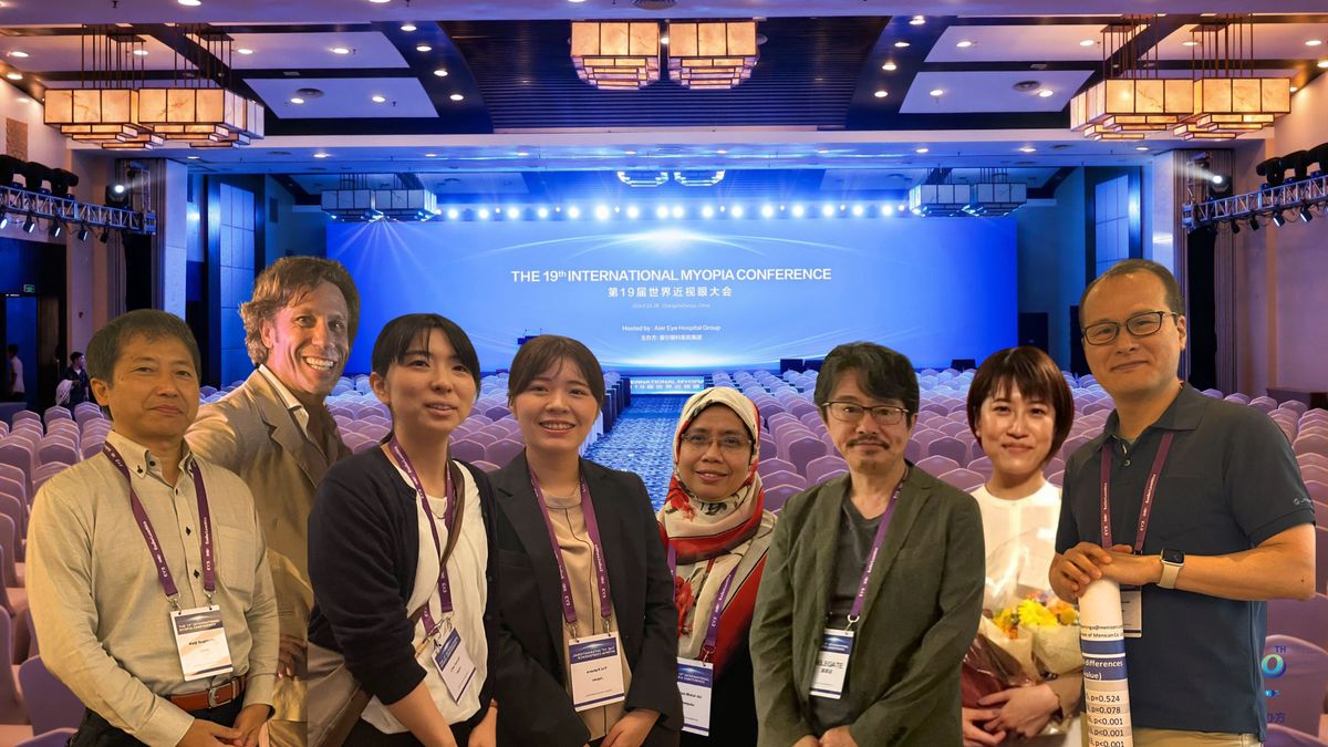
The International Myopia Conference (IMC) 2024 gathered leading experts from the fields of optometry and ophthalmology to explore the most recent advancements and innovations in myopia research. This event provided a platform for sharing cutting-edge findings and fostering collaboration among professionals dedicated to addressing the global myopia epidemic. As part of this, Menicon contributed eight abstracts, showcasing their research in orthokeratology for myopia control. Pictured above are some of the abstract authors, from left to right: Keiji Sugimoto, Dr. Jacinto Santodomingo, Chie Suzuki, Yui Kahara, Bariah Mohd Ali, Takahiro Hiraoka, Hikaru Asai and Asaki Suzaki. In this summary, we highlight key insights from the posters that covered efficacy, safety and potential changes in ocular structures that can occur during ortho-k lens wear.
Eleven years of orthokeratology contact lens wear for slowing myopia progression in children
Authors: Jacinto Santodomingo-Rubido,1 César Villa-Collar,2,3 Ramón Gutiérrez-Ortega,2 Keiji Sugimoto,1 Sachiko Nishimura,1 Steve Newman1
- Menicon Co., Ltd, Nagoya, Japan
- Clínica Oftalmológica Novovision, Madrid, Spain
- Universidad Europea de Madrid, Madrid, Spain
Summary
Myopia typically begins in childhood and can continue progressing well into early adulthood,1 making long-term data on the efficacy of myopia control interventions crucial for understanding their sustained impact over time. This study compared the axial length growth in children wearing ortho-k contact lenses versus single-vision (SV) lenses over an 11-year period. White European children aged 6-12 years with mild to moderate myopia and low astigmatism were assigned to either ortho-k (n = 29) or SV spectacle or contact lenses (n = 24) for two years, with axial length measurements taken every six months. Follow-up measurements were conducted at seven and eleven years from the beginning of the initial 2-year study, with final analysis including data from 10 ortho-k and 10 SV wearers who consistently attended all study visits. The results showed that ortho-k lens wear significantly reduced axial elongation compared to the SV lens group, by 38% (0.693mm total in axial length) after 11 years of wear. These findings suggest that ortho-k lenses are effective in slowing the progression of myopia in children over the long term.
Figure 1: Mean change in axial length (from baseline) ± standard deviation for the orthokeratology and control groups at each time visit. Error bars represent one standard error of the mean.
Abstract
Purpose: To compare axial length growth between a group of orthokeratology contact lens wearers (OK) and a control group of distance, single-vision spectacle lens wearers (CT) over an 11-year period.
Methods: White European subjects 6-12 years-old with myopia -0.75 to -4.00DS and astigmatism ≤1.00DC were prospectively allocated OK or CT for two years. Axial length measurements (Zeiss, IOLMaster) were taken at 6-month intervals during the initial 2 years of the study. Subject were contacted approximately 5 and 9 years later (i.e., 7 and 11 years after the beginning of the study, respectively) and axial length measurements repeated. Changes in axial length (relative to baseline) over an 11-year period were compared between groups using a ‘modified or per-protocol’ intent-to-treat approach whereby data from all subjects, regardless of whether some subjects not attended all study visits or were lost to follow-up at different time points, was analyzed using mixed models with repeated measures to prevent data from subjects being removed from the analysis whenever ≥1 data points were missing (i.e., listwise deletion). Subjects who switched lens wear modalities were excluded from the analysis, but subjects who switched from distance single-vision spectacles to distance single-vision soft contact lenses after the initial two years of the study were included in the CT.
Results: Thirty-one OK and 30 CT subjects were initially recruited; 29 OK and 24 CT completed the initial 2-years study; 10 OK and 15 CT subjects attended the 11-years visit; but only 10 OK and 10 CT subjects attended all study visits. Statistically significant changes in axial length were found over time, between groups and for the time*group interaction (all p≤0.001). Changes over time were statistically significant for all pairs of time points (all p≤0.001), except for the pairs 0.5- vs. 1-, 1- vs. 1.5- and 1.5- vs. 2-years (all p>0.05). In comparison to the CT group, OK lens wear reduced axial elongation [95% confidence intervals] by -0.043mm [-0.102 to 0.016] (-26%, p=0.148), -0.101mm [-0.169 to -0.033] (-31%, p=0.005), -0.143mm [-0.259 to -0.028] (-28%, p=0.016), -0.221mm [-0.365 to -0.076] (-32%, p=0.003),-0.448mm [-0.921 to 0.024] (-33%, p=0.062) and -0.693mm [-1.443 to 0.058] (-38%, p=0.069) following 0.5, 1, 1.5, 2, 7 and 11 years of lens wear, respectively.
Conclusion: OK lens wear provided a substantial slowing in the axial elongation of the eye, with a treatment effect of up to -0.693mm (-38%) following 11 years of lens wear in comparison with single-vision spectacle lens wear. These results provide further support towards the longer-term efficacy of OK in slowing myopia progression in children.
A novel approach for estimating the efficacy of orthokeratology lenses in slowing myopia progression
Authors: Jacinto Santodomingo-Rubido,1 Sin-Wan Cheung,2 César Villa-Collar3 and the ROMIO/MCOS/TO-SEE Groups
- Global R&D, Menicon Co., Ltd, Nagoya, Japan
- School of Optometry, The Hong Kong Polytechnic University, Hung Hom, Kowloon, Hong Kong, China
- Optics & Optometry Department, Faculty of Health Sciences, Universidad Europea, Madrid, Spain
Summary
A key aspect of myopia management in clinical practice involves illustrating the benefits of myopia control interventions and providing estimates on efficacy. This study aimed to estimate the efficacy of ortho-k contact lenses in slowing myopia progression based on axial length data. Pooled data from three prospective studies, involving 101 ortho-k wearers and 88 single-vision (SV) spectacle wearers, were analyzed over a two-year follow-up period. Axial length measurements were taken at baseline and every six months. The mean change in axial length at 24 months was compared between the ortho-k and SV groups, showing a significantly lower axial elongation in the ortho-k group (0.41 mm vs. 0.65 mm, or 0.24mm absolute effect over two years). Additionally, robust correlations found between changes in axial length and mean spherical refractive error (MSE) in the SV group at four different time points (6, 12, 18, and 24 months) were used to estimate refractive myopia progression in the ortho-k group. The study found that ortho-k lenses slowed myopia progression by -0.47 D over two years compared to SV lenses, providing a valuable reference for informing patients about the expected outcomes of ortho-k treatment. In clinical practice, it is also important to note risk factors such as age2 to also provide more tailored insights on myopia control expectations for patients.
Abstract
Purpose: To estimate the efficacy of orthokeratology contact lens wear in slowing myopia progression based on axial length data.
Methods: Pooled data from three prospective studies, which evaluated the use of orthokeratology for slowing the axial elongation in 101 children in comparison to a parallel control group of 88 distance, single-vision spectacle lens wearers who completed a 2-year follow-up period, were used for analysis. Collectively, the pooled mean change in axial length ± standard deviation at the 24-month visit in comparison to baseline for the orthokeratology and single-vision spectacle groups were 0.41 ± 0.25 and 0.65 ± 0.3 mm, respectively, thus orthokeratology lens wear provided an absolute, accumulative treatment effect following 2-years of lens wear of 0.24 mm in comparison to the control group of single-vision spectacles, with a 95% confidence interval of 0.19 to 0.30 mm. Correlations and simple linear regression equations for the relationship between the change in mean spherical refractive error (MSE) and the change in axial length (AL) for the control group of spectacle lens wearers were calculated at four different time points of (i.e., 6-, 12-, 18- and 24-months of lens wear), and these were used for converting changes in AL into changes in MSE in the orthokeratology lens group.
Results. Strong correlations were found between the change in MSE and the change in AL relative to baseline for the control group of spectacle lens wearers at all time points, with the correlation becoming stronger with longer periods of follow-up (i.e., 6-months [r=-0.469, y=-1.4938x+0.0205, p<0.001]; 12-months [r=-0.642, y=-1.6923x+0.0897, p<0.001]; 18-months [r=-0.808, y =-1.8291x+0.032, p<0.001]; and 24-months [r=-0.858, y=-2.0033x + 0.0353, p<0.001]). The estimated mean change in MSE (± standard deviation) at the 24-month visit in comparison to baseline for the orthokeratology was -0.77 ± 0.51 D, whereas that for the control group of distance, single-vision spectacle groups was -1.24 ± 0.71 D. Thus, it is estimated that the orthokeratology arm had a lower increase in MSE than the control by -0.47 D, with a 95% confidence interval of -0.37 to -0.56 D.
Conclusions. A novel approach for estimating the efficacy of orthokeratology lenses in slowing myopia progression is proposed, which estimates that orthokeratology contact lenses reduce the progression of myopia by -0.46 D following 2-years of lens wear in comparison to distance, single-vision spectacle lens wear. These results might be particularly useful for eye care practitioners engaged in fitting orthokeratology lenses for myopia management interested in estimating changes in myopia progression based on axial length data.
Comparison of axial elongation rates before, during, and after orthokeratology
Authors: Takahiro Hiraoka,1 Sayuri Ninomiya,2 Takao Ito3 and Masamichi Kanegae3
- University of Tsukuba, Tsukuba, Japan
- Itami Chuo Eye Clinic, Hyogo, Japan
- Alpha Corporation, Tokyo, Japan
Summary
A rebound effect in myopia control is when myopia progression accelerates after discontinuing a myopia control treatment.3 The concern with rebound effects is that any gains made in slowing myopia progression during treatment could be lost or diminished once the treatment is halted. This study investigated the rate of axial elongation (RAE) before, during, and after ortho-k (OK) treatment to assess the presence of a rebound effect after discontinuation. Retrospective data from 10 patients (18 eyes) were analyzed, with a mean starting age of OK treatment at 11.9 (range 9 to 16) years. The study found that RAE was highest before OK treatment (0.54 mm/year), significantly reduced during OK treatment (0.03 mm/year), and moderately increased after stopping OK (0.30 mm/year). While the mean age at cessation wasn’t specified, longer OK treatment duration and later initiation were associated with slower RAE following cessation. These findings suggest that OK treatment effectively slows axial elongation, with its duration and starting age influencing post-treatment outcomes. It remains essential to monitor patients following the discontinuation of any myopia control intervention to ensure that progression does not resume, especially for younger patients.4
Abstract
Purpose: In 2017, Cho et al. Reported a study (Discontinuation of orthokeratology on eyeball elongation (DOEE). Cont Lens Anterior Eye. 2017;40:82-87) on the rebound phenomenon of axial elongation and myopia progression after discontinuation of orthokeratology (OK). It was found that stopping OK lens wear at or before the age of 14 years led to a more rapid increase in axial length. However, no reports have examined changes in axial length before the initiation of treatment, leaving uncertainty regarding whether a rebound phenomenon truly occurs after OK cessation. Therefore, in this study, we aimed to investigate axial length changes before OK, during OK, and after OK cessation using retrospective data from actual prescriptions to determine the presence of a rebound phenomenon.
Methods: The study included patients who initially wore regular contact lenses or glasses for myopia correction, then underwent OK treatment using α ortho-K lenses (Alpha Corporation, Nagoya, Japan) for a certain period before discontinuation, and continued to visit the same clinic after discontinuing OK treatment. The rate of axial elongation (RAE) was examined during three periods: before OK treatment, during OK treatment, and after OK treatment cessation. Additionally, factors influencing RAE after OK treatment cessation were assessed.
Results: A total of 10 cases (18 eyes) from medical records were selected for the study (2 males, 8 females). The starting age of OK treatment was 11.9±2.1 (mean±SD) years (range: 9 to 16 years), and the spherical equivalent refractive error at the initiation of OK treatment was -2.85±1.76D (range: -5.63 to -0.88D), with astigmatism of -0.52±0.47D (range: -1.75 to 0.00D).
Raes were as follows: before OK treatment, 0.54±0.33mm/year; during OK treatment, 0.03±0.17mm/year; and after OK treatment cessation, 0.30±0.14mm/year. Significant differences were observed among the three groups (p=0.0153, Tukey-Kramer).
RAE after OK treatment cessation showed a significant negative correlation with the duration of OK treatment (r=0.6051, p=0.0130) and the starting age of OK treatment (r=0.6037, p=0.0133).
Conclusions: There were differences in RAE before, during, and after OK treatment cessation. During OK treatment, RAE was the slowest, followed by after OK treatment, and then before OK treatment. No significant rebound phenomenon was observed after OK treatment cessation, and overall, RAE was strongly suppressed during OK treatment. Factors affecting the after OK treatment included the duration of OK treatment and the starting age of OK treatment, with longer treatment durations and later initiation of OK treatment associated with slower RAE after OK discontinuation.
Cleaning efficacy and anti-bacterial properties of contact lens care solutions for orthokeratology lenses
Authors: Yui Kahara,1 Sayaka Goto,1 Jacinto Santodomingo-Rubido,1 Taizo Sumide1
- R&D Center, Menicon Co. Ltd., Kasugai, Aichi, Japan
Summary
Cleaning and safety in ortho-k use often attracts attention due to the potential risk of infections, making proper hygiene and care essential to minimize complications and maintain lens performance.5 This study evaluated the cleaning efficacy and anti-adhesion properties of various contact lens care solutions used on ortho-k lenses, particularly in reducing deposits and minimizing the risk of Pseudomonas aeruginosa (PA) adhesion, which could increase the risk of infection. Ortho-k lenses were subjected to artificial tear film deposits to simulate wear and then cleaned as follows, with PA inoculation after cleaning to test adhesion.
- Group A: Not cleaned (Control 1)
- Group B: Treated with contact lens cleaning/disinfecting solutions
- Group C: Unworn OK lenses (Control 2)
Care solutions in groups B1, B2, and B3 were assessed with and without rubbing.
The results showed that all solutions significantly reduced both lens deposits and PA adhesion, with the chlorine-based solution-cleaned lenses appearing like unworn lenses. The study concluded that daily rubbing, combined with appropriate cleaning solutions (especially chlorine-based) effectively removes deposits and minimizes bacterial adhesion, reducing the risk of lens-related infections. This research helps eye care professionals make informed recommendations on cleaning regimens to enhance the safety and long-term efficacy of ortho-k lens use for their patients.
Figure 2: Total deposition on OK lenses across different treatment conditions. Group A (Control 1) shows 100% normalization of V/P value, representing untreated lenses. Groups B1, B2, and B3 exhibit significant reductions in deposition when lenses are cleaned with contact lens solutions, with or without rubbing. Group C (Control 2) represents unworn OK lenses, showing minimal deposition. Notably, rubbing the lenses (Groups B1, B2, and B3 with rubbing) enhances cleaning efficacy, as indicated by lower V/P values.
Abstract
Purpose: Orthokeratology (OK) contact lenses are effective for slowing myopia progression in children. However, deposits onto OK lenses can be difficult to remove, particularly along the reverse curve area. Increased deposition on OK lenses could potentially lead to a greater risk of contact lens-related infection. Thus, the purpose of this study is to evaluate the cleaning efficacy and anti-adhesion properties against Pseudomonas aeruginosa (PA) of contact lens care solutions on OK lenses in vitro.
Method: OK lenses were soaked in an artificial solution mimicking that of the tear film for 7hr at 80℃. Subsequently, artificially deposited OK lenses were randomly allocated to three groups, including a group that acted as a control in which lenses were not cleaned (Group A); a second group in which lenses were treated with: two different types of multi-purpose solutions (MPS) including a rubbing step: Groups B1 and B2; a hydrogen peroxide solution including a rubbing step: Group B3; and a chlorine-based solution containing sodium hypochlorite without rubbing step: Group B4; and a third group of unworn OK lenses (Group C). One measurement of the cleaning efficacy in removing deposits from OK lenses was taken and quantified as the total volume of cloudiness over the lens surface in terms of volume per unit pixel (V/P) using image analysis software JustTLC (Sweday). After lens cleaning, each OK lens (Group A, Groups B1-B4, and Group C) was inoculated with PA for 4hr at 37℃ and the number of PA adhesion onto the lens surface was quantified by reverse transcriptase-polymerase chain reaction; measurements were made in three lenses per group and a mean was obtained.
Results: Compared with Group A, the V/P value and the adhesiveness of PA for the different OK lens conditions reduced from 100% to 10% and 25%, respectively in Group B1; to 10% and 17%, respectively in Group B2; to 8% and 78%, respectively in Group B3; to 6% and 57%, respectively in Group B4; and to 4% and 50%, respectively in Group C.
Conclusion: The two MPS types (Groups B1 and B2) and the hydrogen peroxide solution (Group B3), which include a rubbing step as part of the care regime, and the chlorine-based solution (Group B4) significantly removed deposits and reduced PA adhesion from the surface of OK lenses. Moreover, the chlorine-based solution (Group B4) was able to remove deposits and reduce PA adhesion to a level like that found in unworn OK lenses (Group C). These results indicate that daily rubbing of OK lenses prior to using a cleaning/disinfecting solution and the regular use of chlorine-based solution are effective in removing deposits and keeping lenses clean, thus minimizing the adhesion of PA onto the lens surface, ultimately contributing to a reduced risk of OK lens-related complications.
Effect of coexistence with bacteria treated with multipurpose solution on excystment of Acanthamoeba castellanii
Authors: Chie Suzuki,1 Mayo Otsuka,1 Jacinto Santodomingo-Rubido,1 Taizo Sumide1
- R&D Center, Menicon Co. Ltd., Kasugai, Aichi, Japan
Summary
Acanthamoeba excystment is the process by which Acanthamoeba cysts, which are dormant, highly resistant forms of the amoeba, transform back into their active, feeding form called trophozoites. This process typically occurs when environmental conditions become favorable, such as the presence of nutrients or bacteria.6 Excystment is significant in the context of contact lens wear because once Acanthamoeba trophozoites are active, they can adhere to the cornea and cause serious eye infections, such as Acanthamoeba keratitis. This study investigated the potential for Acanthamoeba excystment to occur in the presence of bacteria found in ortho-k lens cases. Acanthamoeba cysts were exposed to bacteria such as Pseudomonas rhodesiae and Streptococcus salivarius, with results showing that the cysts' excystment rates increased significantly in their presence. Additional experiments revealed that while UV-treated bacteria could still induce excystment, bacteria treated with a multipurpose solution (MPS) did not. The findings suggest that while basic bacterial elimination may not fully prevent Acanthamoeba-related infections, more rigorous disinfection protocols can support safer ortho-k lens wear. Clinicians should therefore emphasize disinfection and hygiene protocols at every visit to ensure patients understand the importance of proper lens care.
Abstract
Aim: Orthokeratology lenses are becoming a relatively common method for slowing myopia progression in children. To ensure safe contact lens wear, it is important to minimize the risk of any contact lens-related infection. Although rare, Acanthamoeba keratitis is serious ocular infection that could occur with orthokeratology contact lens wear. Previous studies have shown that Acanthamoeba castellanii trophozoite proliferate in coexistence with bacteria, and excystment of cyst could also be induced in coexistence with bacteria. This study investigated whether Acanthamoeba excystment is induced by its coexistence with isolates found in the lens cases of orthokeratology lens wearers or by bacteria treated with ultraviolet light (UV) and multipurpose solution (MPS).
Methods: A.castellanii ATCC 50370 cysts, together with coexisting bacteria Pseudomonas rhodesiae and Streptococcus salivarius isolated from orthokeratology lens cases, were used in experiment I, and Escherichia coli and Streptococcus epidermidis were used in experiment I and II. In experiment I, cysts in a concentration of 105cells/mL were inoculated with each bacterial suspension in a concentration of approximately 102 to 107CFU/mL. Subsequently, trophozoites and cysts were stained with calcofluor white solution, and counted under a fluorescence microscope. In experiment II-A, a suspension of E.coli or S.epidermidis in a concentration of approximately 106 to 107CFU/mL was treated with UV, and subsequently inoculated with cysts in a concentration of 105cells/mL. In experiment II-B, a suspension of E.coli or S.epidermidis in a concentration of approximately 107CFU was mixed with a MPS (MeniCare Plus, Menicon Co., Ltd) for 24 hour, collected by centrifugation, and subsequently inoculated with cysts in a concentration of 105cells/mL. Subsequently, trophozoites and cysts were stained and counted as in experiment I. A suspension with no inoculum was used as control in all experiments.
Results: In experiment I, excystment rates increased by approximately 2- to 3-fold and 4- to 12-fold, respectively in the inoculum containing P.rhodesiae and S.salivarius in comparison to the control. Additionally, excystment rates of the suspension containing E.coli and S.epidermidis increased approximately 4- to 6-fold and 2- to 4-fold, respectively in comparison to the control. In experiment II-A, excystment rates of the E.coli treated with UV increased by approximately 3-fold, and S.epidermidis treated with UV increased by approximately 5-fold in comparison to control. In experiment II-B, excystment rates of the both bacteria treated with MPS were comparable to control.
Conclusion: The results of this study show that Acanthamoeba excystment can be induced when cysts coexist with isolates from orthokeratology lens cases. Furthermore, the results also indicate that although bacteria treated with UV could also aid in inducing excystment, that’s unlikely to be the case when bacteria is treated with MPS. Taken together, the results of this study suggest that to ensure safe and successful orthokeratology lens wear killing bacteria might not be enough to reduce the risk of ocular infection, and thus greater levels of disinfection should be considered.
Effect of ortho-k lenses on cornea endothelial cells, cornea thickness, and ocular surface in myopic children
Authors: Yu Chen Low,1 Bariah Mohd-Ali,1 Mizhanim Mohamad Shahimin,1 Hamzaini Abdul Hamid,2 Wan Haslina Wan Abdul Halim3
- Optometry and Vision Science Program, Research Centre for Community Health, Faculty of Health Science, Universiti Kebangsaan Malaysia
- Department of Radiology, Faculty of Medicine, Universiti Kebangsaan Malaysia
- Department of Ophthalmology, Faculty of Medicine, Universiti Kebangsaan Malaysia.
Summary
This study evaluated the impact of ortho-k treatment on corneal endothelial cell morphology, corneal thickness, and ocular surface health in myopic children over 12 months, compared to SV spectacle lens wearers. Seventy children aged 8-9 years were recruited, with 45 fitted with ortho-k and 25 with SV spectacles. Measurements of corneal endothelial cells and ocular health were taken at baseline and 12 months using specular microscopy and standard slit lamp examinations. The results showed no significant differences between the ortho-k and SVS groups in endothelial cell density, hexagonality, or coefficient of variation. The ortho-k group showed a small but statistically significant reduction in central corneal thickness (CCT) of 4 microns over 12 months, as expected due to the lens influence on corneal topography, while the SVS group showed no change. Some minor complications were reported in the ortho-k group, including styes and conjunctivitis, but no significant corneal staining or severe complications were noted between the groups. The findings suggest that ortho-k is a safe option for myopia control in children, though ongoing monitoring and proper lens management are essential.
Abstract
Purpose: To investigate changes in cornea endothelial cells morphologies, cornea thickness, and ocular surface in myopic children undergoing orthokeratology (OK) treatment for 12 months using specular microscopy. The results were compared to single vision spectacle wearers (SVS).
Patients and Methods: 70 children with myopia (aged 8–9 years old) were recruited. A total of 45 children were fitted with OK, and 25 were fitted with SVS. Corneal endothelial cell morphologies (ECD, COV, HEX, and CCT) were measured by using a non-contact specular microscope (SP-3000P; Topcon, Japan). Standard slit lamp investigations were conducted using a binocular slit lamp (Righton MW50D LED, Tokyo, Japan) to evaluate the anterior segment including cornea, bulbar and tarsal conjunctiva (upper and lower), tear film quality, and photographed recording of the subject’s eye and OK lens fitting. All the measurements were conducted at the baseline and 12 months.
Results: When comparing between groups, no sig differences were shown in the ECD, HEX and COV in OK group and SVS group after 12 months of treatment (p>0.05). However, the mean difference in CCT showed a statistically sig reduction in the OK group at 12 months when compared to baseline. In the OK group, CCT decreased from 528.53 ± 22.26 at the baseline to 524.87 ± 23.15 at 12 months after treatment (p = 0.032). Whereas in the SVS group, the CCT remained at the same value and showed no significant difference at 12 months, (p = 0.767). After 12 months of OK lens wear, 5 subjects reported to have stye, 1 subject reported to have foreign body (FB) sensation, 1 subject reported to have contact lens peripheral ulcer (CLPU) and 2 subjects reported to have conjunctivitis. When comparing between SVS and OK group, no sig complications and corneal staining between SVS group and OK group after 12 months all (p>0.05).
Conclusion: Ortho-K treatment, therefore, is a safe option for myopia control in the children population. However, close monitoring for contact lens compliance, appropriate clinical standards in lens fitting, and patient management are recommended.
Comparison of peripheral eye length measurements between Orthokeratology and single vision spectacle lens wearers, using MRI analysis
Authors: Bariah Mohd-Ali,1 Low Yu-Chen,1 Mizhanim Mohamad Shahimin,1 Hamzaini Abdul-Hamid,2 Siti Salasiah Mokri,3 Norhani Mohidin1
- Optometry and Vision Science Program, Research Centre for Community Health, Faculty of Health Science, Universiti Kebangsaan Malaysia
- Department of Radiology, Faculty of Medicine, Universiti Kebangsaan Malaysia
- Department of Ophthalmology, Faculty of Medicine, Universiti Kebangsaan Malaysia.
Summary
Traditionally, central axial length has been the primary measurement used for determining myopia progression, as it directly correlates with the degree of myopia. However, peripheral eye length (PEL) is gaining recognition as an important factor, as it provides insights into how the eye grows in different regions, not just centrally. This study examined changes in PEL in myopic children undergoing ortho-k treatment for 12 months compared to those wearing SV spectacles, using MRI. Seventy children aged 8-9 years were included, with 45 fitted with ortho-k lenses and 25 with SV. MRI measurements of PEL and axial length (AL), along with peripheral refraction (PR) at various angles, were taken at baseline and after 12 months. The results showed that the SV group experienced more elongation of both PEL and AL across all eccentricities compared to the ortho-k group. The ortho-k group exhibited significant PEL elongation mainly beyond 20 degrees, while central AL and closer eccentricities showed reductions. PR in the ortho-k group became more myopic, whereas the SV group showed hyperopic shifts. The study suggests that as myopia progresses with ortho-k wear, ortho-k provide myopic defocus and also it causes the eye to grow in a way that promotes peripheral myopic defocus through the elongation of PEL over AL. This potentially provides greater understanding of the myopia control mechanism behind ortho-k and how the eye responds to defocus.
Abstract
Purpose: To investigate changes in peripheral eye length (PEL) in myopic children undergoing orthokeratology (Ortho-K) treatment for 12 months using MRI. The results were compared to single vision spectacle wearers (SVS).
Methods: 70 children with myopia (aged 8–9 years old) were recruited. A total of 45 children were fitted with Ortho-K, and 25 were fitted with SVS. The PEL and axial length (AL) were measured by using MRI 3-Tesla, whereas central and peripheral refraction (PR) measurements were conducted at ±30 degrees horizontally with nasal (N) and temporal (T) intervals of 10°, 20°, and 30° and with an open field autorefractometer (WAM-5500 Grand Seiko). All the measurements were conducted at the baseline and 12 months.
Results: The MRI analysis indicates that at 12 months, the SVS group showed more elongation of the PEL and AL at all eccentricities than the Ortho-K group did (p < 0.05). The Ortho-K group only showed significant PEL elongation beyond 20 degrees at N20, N30, T20, and T30 (p < 0.05); however, a significant reduction in the AL was detected in the center AL, N10, and T10 (p < 0.05). All eccentricities in the relative PR of the Ortho-K group were significantly more myopic than at the baseline (p < 0.001), whereas in the SVS group, all eccentricities in the relative PR were shown to be significantly more hyperopic than at the baseline (p < 0.001). The PEL and PR showed negative correlations at 12 months in the Ortho-K group.
Conclusions: MRI analysis can be utilized to describe changes in PEL in myopic children. It appears that as myopia progressed in Ortho-K lens wearers, the PEL increased by a greater amount than the AL did; thus, the retina was reshaped to become increasingly oblate and to display peripheral myopic defocus.
Factors associated with axial length and subfoveal choroidal thickness in myopic children
Authors: Hikaru Asai1,2, Yukiko Okabe3, Asaki Suzaki1, Keiji Sugimoto1, Shinjiro Kono2, Ai Shibata2, Sayaka Yamao2,3, Motohiro Kamei2, Atsuya Miki2,3
- Menicon Co., Ltd.
- Aichi Medical University
- Aichi Medical University Eye Center
Summary
Subfoveal choroidal thickness (SfChT) is gaining traction in myopia research because changes in choroidal thickness have been linked to alterations in eye growth and myopia progression.7 This study aimed to identify factors associated with axial length and SfChT in myopic Japanese children. Fourteen children aged 8 to 10 years were assessed before starting ortho-k, with a mean spherical equivalent (SE) of -1.93 ± 1.03 D and an average axial length of 24.41 ± 0.81 mm. The mean SfChT was 248.4 ± 58.8 µm. Multivariate analysis showed that longer axial length was significantly associated with male gender, more myopic SE, smaller corneal refraction, and thinner lens thickness. In contrast, SfChT was primarily correlated with lens thickness, showing no direct association with axial length. These findings suggest that while axial length and choroidal thickness are influenced by different factors, understanding these associations allows clinicians to more closely monitor those with factors associated with longer axial length.
Figure 3: Table 3 presents the regression coefficients (β) and corresponding p-values for associations with AL and SfChT. Significant predictors for AL include gender (p = 0.0215), spherical equivalent (SE; p < 0.0001), and average keratometry (Average K; p < 0.0001). For SfChT, only LT showed a significant relationship (p = 0.0157).
Abstract
Purpose: Previous studies suggested an association between the choroidal thickness and the progression of myopia. The aim of this study is to investigate and identify factors associated with axial length and subfoveal choroidal thickness of myopic children in Japan.
Method: Demographic and ocular biometric data were collected from fourteen Japanese children prior to the commencement of orthokeratology treatment at Aichi Medical University Eye Center. Axial length was measured using SS-OCT biometer (ARGOS®, Alcon) and subfoveal choroidal thickness (SfChT) was measured using SS-OCT (OCT-S1, Canon). The software of OCT-S1 was utilized for auto segmentation and measurement of choroidal thickness. Multivariate linear regression analyses were employed to examine the correlation of axial length and SfChT with gender, age, spherical equivalent (SE), corneal refraction, lens thickness and intraocular pressure (IOP).
Results: The age of the participants was 9.5±1.0 years old (mean±standard deviation, range : 8 to10 years old). SE was -1.93±1.03 D (range : -0.50 to-4.50 D), axial length was 24.41±0.81 mm (range : 22.72 to26.10 mm), and SfChT was 248.4±58.8 µm (range : 144 to364 µm). Axial length was significantly correlated with gender (male>female, p=0.0013), SE (β=-0.40, p<0.0001), corneal refraction (β=-0.53, p<0.0001) and lens thickness (β=-0.95, p=0.0141). SfChT was correlated with lens thickness only (β=205.07, p=0.0037).
Conclusions: In myopic children, longer axial length was associated with male gender, more myopic SE, smaller corneal refraction and thinner lens. Axial length was not associated with SfChT. Thicker SfChT was associated with thicker lens. These findings may provide further insight towards the potential factors that may contribute to the variation of the choroidal thickness.
Meet the Authors:
About Jeanne Saw
Jeanne is a clinical optometrist based in Sydney, Australia. She has worked as a research assistant with leading vision scientists, and has a keen interest in myopia control and professional education.
As Manager, Professional Affairs and Partnerships, Jeanne works closely with Dr Kate Gifford in developing content and strategy across Myopia Profile's platforms, and in working with industry partners. Jeanne also writes for the CLINICAL domain of MyopiaProfile.com, and the My Kids Vision website, our public awareness platform.
This content is brought to you thanks to an educational grant from
References
- COMET Group. Myopia stabilization and associated factors among participants in the Correction of Myopia Evaluation Trial (COMET). Invest Ophthalmol Vis Sci. 2013 Dec 3;54(13):7871-84.
- Donovan L, Sankaridurg P, Ho A, Naduvilath T, Smith ELI, Holden BA. Myopia progression rates in urban children wearing single-vision spectacles. Optom Vis Sci. 2012;89:27-32.
- Sánchez-Tena MÁ, Ballesteros-Sánchez A, Martinez-Perez C, Alvarez-Peregrina C, De-Hita-Cantalejo C, Sánchez-González MC, Sánchez-González JM. Assessing the rebound phenomenon in different myopia control treatments: A systematic review. Ophthalmic Physiol Opt. 2024 Mar;44(2):270-279.
- Gifford KL, Richdale K, Kang P, Aller TA, Lam CS, Liu YM, Michaud L, Mulder J, Orr JB, Rose KA, Saunders KJ, Seidel D, Tideman JWL, Sankaridurg P. IMI - Clinical Management Guidelines Report. Invest Ophthalmol Vis Sci. 2019 Feb 28;60(3):M184-M203.
- Bullimore MA, Johnson LA. Overnight orthokeratology. Cont Lens Anterior Eye. 2020 Aug;43(4):322-332.
- Khunkitti W, Lloyd D, Furr JR, Russell AD. Acanthamoeba castellanii: growth, encystment, excystment and biocide susceptibility. J Infect. 1998 Jan;36(1):43-8.
- Liu Y, Wang L, Xu Y, Pang Z, Mu G. The influence of the choroid on the onset and development of myopia: from perspectives of choroidal thickness and blood flow. Acta Ophthalmol. 2021 Nov;99(7):730-738.
Enormous thanks to our visionary sponsors
Myopia Profile’s growth into a world leading platform has been made possible through the support of our visionary sponsors, who share our mission to improve children’s vision care worldwide. Click on their logos to learn about how these companies are innovating and developing resources with us to support you in managing your patients with myopia.


